WEEK 24
Part
B
Secondary Endpoint
WEEK 52
Part
B-C
Secondary Endpoint
Erefs total score was reduced at weeks 24 and 52
Thresholds for clinically meaningful changes in EREFS scores have not been established. Additionally in Part B, this endpoint was ordered after the point at which hierarchical testing procedure failed. Results are descriptive in the extended active treatment period at Week 52. Definitive conclusions cannot be made due to limitations associated with extended active treatment design, including lack of comparator arm and decreasing sample size.
EREFS is a clinician-reported rating of the severity of 5 endoscopic features:
edema, rings, exudates, furrows, and stricture
EDEMA
(Grades 0-2)
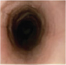
Grade 1
RINGS
(Grades 0-3)
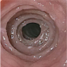
Grade 3
EXUDATES
(Grades 0-2)
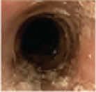
Grade 2
FURROWS
(Grades 0-2)
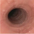
Grade 2
STRICTURE
(Grades 0-2)

Grade 1
Scores from the proximal and distal regions range from 0 to 9.3
The EREFS total score is the sum of those 2 regions (range 0-18).6
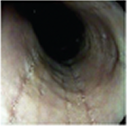
E1, R1, E0, F1, S0
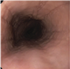
E1, R0, E0, F2, S0
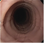
E1, R2, E0, F1, S1
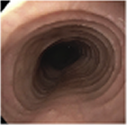
E1, R2, E1, F1, S0
a Endoscopic images are from a region of the esophagus in different patients and are not composite EREFS total scores. Endoscopic images are for reference only.
Adapted by permission from BMJ Publishing Group Limited. Gut, Hirano I et al, vol. 62, pages 489-495 (2013). Adapted with permission from Wolters Kluwer Health, Inc.: Kia L, Hirano I. Advances in the endoscopic evaluation of eosinophilic esophagitis, Curr Opin Gastroenterol, vol. 32(4), pages 325-331. https://journals.lww.com/co-gastroenterology/ Reprinted from J Allergy Clin lmmunol Pract, vol. 6(5), Steinbach EC et al, Eosinophilic esophagitis and the eosinophilic gastrointestinal diseases: approach to diagnosis and management, pages 1483-1495 (2018), with permission from Elsevier.